
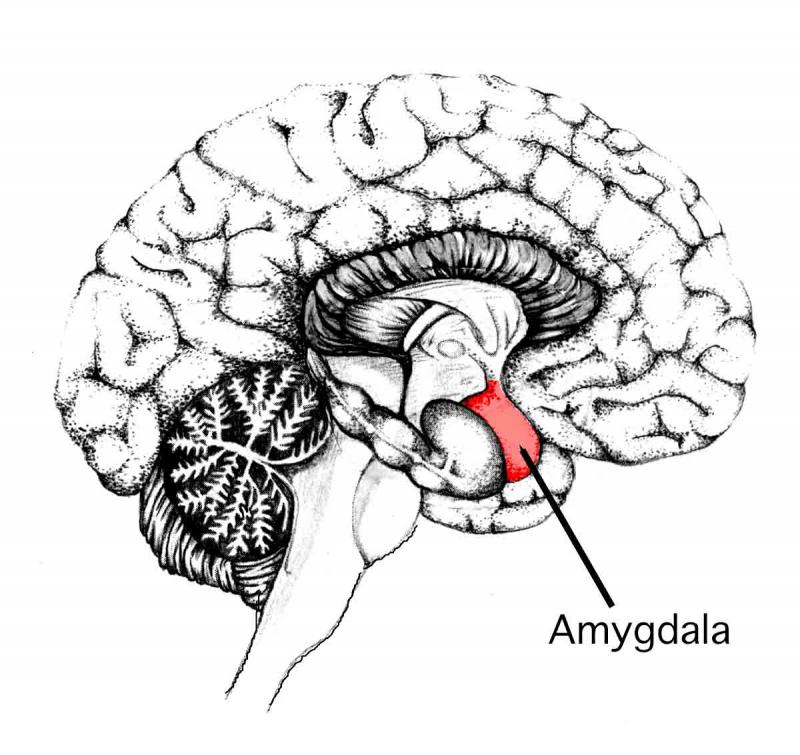
The amygdala (Latin, corpus amygdaloideum) is an almond-shape set of neurons located deep in the brain's medial temporal lobe.
Shown to play a key role in the processsing of emotions, the amygdala forms part of the limbic system.
In humans and other animals, this subcortical brain structure is linked to both fear responses and pleasure.
Its size is positively correlated with aggressive behavior across species.
In humans, it is the most sexually-dimorphic brain structure, and shrinks by more than 30% in males upon castration.
Conditions such as anxiety, autism, depression, post-traumatic stress disorder, and phobias are suspected of being linked to abnormal functioning of the amygdala, owing to damage, developmental problems, or neurotransmitter imbalance.
Source:
The above post is reprinted from materials provided by Massachusetts General Hospital. Note: Materials may be edited for content and length.
In humans and other animals, this subcortical brain structure is linked to both fear responses and pleasure.
Its size is positively correlated with aggressive behavior across species.
In humans, it is the most sexually-dimorphic brain structure, and shrinks by more than 30% in males upon castration.
Conditions such as anxiety, autism, depression, post-traumatic stress disorder, and phobias are suspected of being linked to abnormal functioning of the amygdala, owing to damage, developmental problems, or neurotransmitter imbalance.
 | ||||||
| credit: brynmawr.edu |
Structure deep within the brain may contribute to a rich, varied social life
Massachusetts General Hospital,
Scientists
have discovered that the amygdala, a small almond shaped structure deep
within the temporal lobe, is important to a rich and varied social life
among humans. The finding was published this week in a new study in Nature Neuroscience
and is similar to previous findings in other primate species, which
compared the size and complexity of social groups across those species.
"We
know that primates who live in larger social groups have a larger
amygdala, even when controlling for overall brain size and body size,"
says Lisa Feldman Barrett, PhD, of the Massachusetts General Hospital
(MGH) Psychiatric Neuroimaging Research Program and a Distinguished
Professor of Psychology at Northeastern University, who led the study.
"We considered a single primate species, humans, and found that the
amygdala volume positively correlated with the size and complexity of
social networks in adult humans."
The researchers also performed an exploratory analysis of all the subcortical structures within the brain and found no compelling evidence of a similar relationship between any other subcortical structure and the social life of humans. The volume of the amygdala was not related to other social variables in the life of humans such as life support or social satisfaction.
"This link between amygdala size and social network size and complexity was observed for both older and younger individuals and for both men and women," says Bradford C. Dickerson, MD, of the MGH Department of Neurology and the Martinos Center for Biomedical Research. "This link was specific to the amygdala, because social network size and complexity were not associated with the size of other brain structures." Dickerson is an associate professor of Neurology at Harvard Medical School, and co-led the study with Dr. Barrett.
The researchers asked 58 participants to report information about the size and the complexity of their social networks by completing standard questionnaires that measured the total number of regular social contacts that each participant maintained, as well the number of different groups to which these contacts belonged. Participants, ranging in age from 19 to 83 years, also received a magnetic resonance imaging brain scan to gather information about the structure of various brain structures, including the volume of the amygdala.
A member of the the Martinos Center at MGH, Barrett also notes that the results of the study were consistent with the "social brain hypothesis," which suggests that the human amygdala might have evolved partially to deal with an increasingly complex social life. "Further research is in progress to try to understand more about how the amygdala and other brain regions are involved in social behavior in humans," she says. "We and other researchers are also trying to understand how abnormalities in these brain regions may impair social behavior in neurologic and psychiatric disorders."
Co-Authors of the Nature Neuroscience paper are Kevin C. Bickart, Boston University School of Medicine; and Christopher I. Wright, MD, PhD, and Rebecca J. Dautoff of the MGH Psychiatric Neuroimaging Research Program and the Martinos Center. The study was supported by grants from the US National Institutes of Health and the US National Institute on Aging.
Source:
The above post is reprinted from materials provided by Massachusetts General Hospital. Note: Materials may be edited for content and length.
Brain Chemical Plays Critical Role In Drinking And Anxiety
University of Illinois at Chicago,
A
brain protein that sustains nerve cells also regulates anxiety and
alcohol consumption in rats, researchers from the University of Illinois
at Chicago report in a study in the Aug. 9 issue of the Journal of
Neuroscience.
In previous studies, the UIC researchers had first identified a gene that controls anxiety and alcohol consumption.
"We knew that gene, called CREB, controls the expression of a number of important genes in the brain," said Dr. Subhash Pandey, professor of psychiatry and anatomy and cell biology at UIC and Jesse Brown VA medical center and lead author of the paper. In the new study, they showed that a protein made by one of those CREB-controlled genes affects anxiety and drinking behavior depending on its level in two areas of the brain.
Pandey and his colleagues injected DNA of complementary sequence to the gene of the protein, called brain-derived neurotrophic factor (BDNF), into the brains of rats to block the gene from expressing BDNF. The "anti-sense" DNA was injected into three areas of the amygdala, an area of the brain associated with emotion and fear.
The researchers found that when levels of BDNF in the central and medial areas of the amygdala were lowered, anxiety and alcohol consumption increased. Decreased levels of BDNF in the third area, called the basolateral amygdala, had no effect.
When levels of BDNF in the central and medial amygdala were restored to normal by injecting BDNF, anxiety and alcohol consumption diminished.
The researchers measured anxiety by observing the rat's exploratory behavior in a maze. Alcohol consumption was measured by offering the animals one drinking bottle with water and one with alcohol, and noting the proportion of alcohol imbibed.
BDNF plays a vital role in the growth and maintenance of neurons. Many human studies have suggested that variations in the BDNF gene may be associated with alcoholism and anxiety disorders, Pandey said.
"In people, alcoholism is very frequently associated with anxiety disorders," he said. "And it is well established that high levels of anxiety promote alcohol consumption and also play a crucial role in relapse to alcohol drinking."
Pandey said the new research may suggest a target for drugs to treat or prevent anxiety and alcoholism.
"Our study suggests a molecular, neurochemical mechanism in the amygdala which may be responsible for the association of high levels of anxiety with excessive alcohol-drinking behavior," he said.
Huaibo Zhang, Adip Roy and Kaushik Misra of UIC co-authored the study. The research was supported by grants from the National Institute on Alcohol Abuse and Alcoholism and the Department of Veteran Affairs.
Source:
The above post is reprinted from materials provided by University of Illinois at Chicago. Note: Materials may be edited for content and length.
Getting on 'the GABA receptor shuttle' to treat anxiety disorders
RIKEN,
There
are increasingly precise molecular insights into ways that stress
exposure leads to fear and through which fear extinction resolves these
fear states. Extinction is generally regarded as new inhibitory
learning, but where the inhibition originates from remains to be
determined. Gamma-aminobutyric acid (GABA), the primary inhibitory
chemical messenger in the brain, seems to be very important to these
processes.
A new article in Biological Psychiatry
examined whether during the extinction of fear learning, GABA receptors
may be inserted into the cell surface to reduce the excitability of the
amygdala. Researchers inactivated a protein that links GABAA receptors
to the cell surface. They found that this protein prevented fear
extinction training and the local application of NMDA from increasing
the number of GABAA receptors on the cell surface and enhancing the
inhibition of amygdala nerve cells.
Lin and colleagues show that during fear conditioning, the number of GABAA receptors on the surface of neurons in the amygdala decreases, reducing the extent of inhibition of the neurons in this brain "fear center." When fear is extinguished by dissociating fear cues from unpleasant stimuli, the number of GABAA receptors on the cell surface of the amygdala neurons increases.
How does this happen? The study provides evidence of molecular mechanisms that shuttle GABAA receptors to the cell surface during extinction. The researchers showed that by inactivating a protein involved in the localization of GABAA receptors in the amygdala, they prevented the recruitment of GABA-mediated inhibition and extinction of fear. Dr. John Krystal, Editor of Biological Psychiatry comments: "This research provides evidence that we are starting to untangle the molecular mechanisms through which our cognitive and behavioral therapies might alter brain function."
wow. boring
ReplyDeletesorry my sister did that
Delete